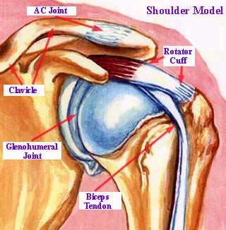
Home
About Us
Contact Us
Find a Doctor
Arthritis Osteoarthritis
Arthritis Types
Rheumatoid Arthritis
Psoriasis
Fractures Fractures
Fracture Types
Adult Fractures
Kids' Fractures
Salter-Harris Fracture
Salter Type I Fx
Salter Type II Fx
Salter Type III Fx
Salter Type IV Fx
Hand & Wrist Wrist Fractures
Scaphoid Fractures
Scaphoid Fx Diag.
Scaphoid Fx. Treat.
Knee Knee Joint Pain
Knee Sprain
Knee Cartilage Tears
Knee Arthroscopy
Knee ACL Surgery
Foot & Ankle Ankle Pain
AnkleFracture
Treating Ankle Sprains
Ankle Braces
Heel Pain
Heel Pain Treatment
Hip Hip Fracture
Hip Pain
Back & Neck Low Back Pain
Spinal Stenosis
Shoulder Shoulder Pain
Shoulder Anatomy
Shldr Impingement
Rotator Cuff Tear
Cuff Tear Symptoms
Shldr Arthroscopy
Shoulder labral tear
Joint Replacement Joint Replacement
Shoulder Replacement
Anesthesia Anesthesia Types
Epidural Anesthesia
Fem. Nerve Block
Interscalene Block
Suprascapular Block
Medications & Treatments Joint Pain Remedies
NSAIDs
Alternative Pain Rx
Pt. Advocacy Patient Advocate
Web Friends

Shoulder Joint Anatomy


Let’s talk about shoulder joint anatomy. As we briefly discussed on the introductory page, it is a ball and socket joint configuration.

The humeral head is the “ball" and the glenoid is the “socket”. Other structures that serve to make up the “shoulder girdle” include the clavicle (collar bone), the acromion (top part of the scapula, or, shoulder blade) and the coracoid process (part of the front portion of the glenoid and an attachment point for ligaments).


The picture above shows some shoulder structures as seen from the front.

What sets the shoulder joint apart from other ball and socket joints, such as the hip, is mainly that the glenoid (socket) is rather shallow and does not, by itself, contribute too much to stability. It also relies upon the integrity of such structures as muscles, tendons and ligaments to maintain that stability and to keep it “in joint”.


This view, from behind, shows more of the muscles and tendon structures that make up the rotator cuff complex, namely the supraspinatus, infraspinatus, and teres tendons.

It also shows the sub-acromial bursa sac, which has a reputation for being a troublesome character. This bursa can become severely inflamed and restrict motion when part of a condition called shoulder impingement

Looking at these pictures, you can see the structures and how they are located either in the front, or the rear of the shoulder.

You can also see how the combination of these muscles, tendons and ligaments work together to not only stabilize the joint, but to provide a wide range of functions and movements.


The glenoid (socket) is, by nature, quite shallow, so a structure surrounds it, called the glenoid labrum, which acts to help “deepen” the socket to increase stability.

This labrum is the source for several types of injuries that can affect shoulder stability, such as the labral tear, and a condition known as the S.L.A.P. tear. More on that later...

Leave Shoulder Joint Anatomy; return to Shoulder Pain

Leave Shoulder Joint Anatomy; returnt to Home Page


Ken Chisholm, Founder/Publisher

"We hope you enjoy your journey through Bone and Joint Pain.com"


Visit OTCOnly.com!


Medical Supplies